Natural zinc concentration in dentine mapped across entire teeth
Researchers in Germany have mapped how the element zinc is distributed throughout teeth in unprecedented detail. The findings could help clinicians better understand how zinc-containing dental materials interact with natural tooth tissue.
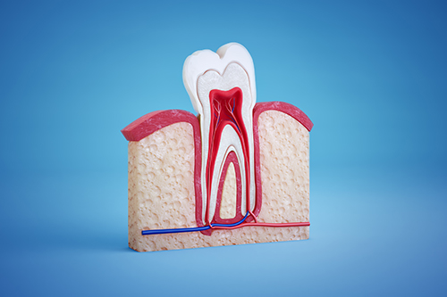
Dentine density and tubule porosity vary markedly across the tooth, especially towards the pulp, and although zinc was already known to increase in this direction, it had not been systematically quantified across entire intact teeth or directly related to these structural gradients.
To investigate how chemistry relates to dentine structure and establish a baseline for comparison in future studies of disease and restorative treatment, the researchers combined micro-CT with quantitative micro-X-ray fluorescence imaging and used healthy, intact teeth. The CT scans provided a 3D map of dentine density and dentinal tubule distribution, while the fluorescence technique measured the concentration of elements such as calcium, phosphorus and zinc throughout the tooth.
The results showed that calcium and phosphorus were distributed relatively uniformly across the tissue. Zinc, however, followed a striking gradient. Its concentration increased fivefold to tenfold from the denser outer regions of dentine towards the pulp. The finding that zinc concentration rose as dentine density decreased suggests that the element is localised in or around the dentinal tubules.
The findings could thus have implications for clinical dentistry. Since zinc is widely used in dental materials, understanding the natural distribution of zinc in dentine may help researchers evaluate how such materials interact with tooth tissue and whether they influence enzymatic processes involved in dentine degradation, with possible implications for bond durability.


